Bichat, XavierAntiquariat.de
5 817 résultats
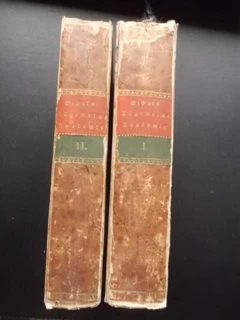
1802réf. 307703ABLeipzig, Crusius, 1802/1803. 8°. XVI, 324 S.; XII, 379 S.; XXII, 343 S.; X, 302 S. Original-Pappbände d. Zt. 2 Bände cplt. (Einbände berieben u. bestoßen, Rücken oben mit Fehlstelle, sonst guter Zustand).
1890réf. 72311890 demi-reliure havane (half binding) grand in-octavo, dos long orné de filet et de filets perlés - titre frappé or (spine without raised band illuminated with a gilt line and with a gilt beaded line and gilt title), plats frottés - papier marbré aux plats (rubbed cover with marbled paper), tranches lisses (smooth edges), illustrations : 455 figures in texte en partie coloriées (pictures in text partly colored), naissances de rousseurs (beginning of the redness marks), 1064 pages, 1890 à Paris Librairie J.-B. Baillière et Fils,
1896Un volume in folio grande (27x38 cm) di 96 pagine con 100 disegni anatomici (molti a piena pagina) su carta pesante. Qualche fioritura. Tutta tela editoriale -- First Edition. An elaborate art manual for sculptors, painters, illustrators and naturalists: is the first such attempt at an anatomy of animals fron the art standpoint.
réf. 2037P., Baillière, sans date (1865), un volume in folio relié en pleine soie bleue marine, étiquette de titre en maroquin rouge (reliure postérieure), (2), 25 PLANCHES
réf. 7494P., Masson, 1894, un volume in folio relié en pleine toile éditeur, titre en lettre dorée au dos et sur le premier plat de couverture (reliure de l'époque), (cachet de bibliothèque sur le faux-titre annulé et ex-libris gratté), 8pp., 48pp., 37 planches dont 2 EN COULEURS
1847réf. MEDI1015Wien, Wallishausser 1847. XVI, 523 S.; XII, 427 S.; Interimsbrosch., nur vord. Umschl. v. Bd. 2 vorhand., dieses händisch beschriftet, randrissig, stockfl. u. verstaubt, tlw. in Lagen gebrochen, ungleich geschnitt., starke Staubränder u. tlw. etw. randrissig, innen guter, sauberer Zustand. Erstausgabe. - HYRTL, Josef. (1810-1894), österreichischer Anatom. Dieses, sein Hauptwerk wurde weltweit zu einem der wichtigsten Lehrbücher an medizinischen Schulen.
1963In-4 gr., lussuosa legatura editoriale in piena pelle (abrasioni alle cerniere), dorso a cordoni, astuccio con cuffia in pelle, 54 cc.nn., con il bellissimo frontespizio figurato e 8 grandi tavole anatomiche di cui 1 a colori. Riproduzione in facsimile della seconda edizione (1493) del primo testo di anatomia con figure originali. E' l'anatomia di Mondino de' Luzzi (1270-1326), il primo insigne anatomista degno di questo nome, pubblicata per la prima volta a Venezia nel 1491 dal medico Giovanni de Ketham, alemanno, nel suo cosiddetto "Fascicolo di medicina". Cfr. Castiglioni "Storia della Medicina", pp. 340, 420. Bella edizione su carta a mano, in tiratura di 350 esemplari numerato. Ben conservato.
1945L. Testut Anatomia Umana 12 volumi (8 tomi). , U.T.E.T. 1945, Legatura artigianale molto bella, ben fatta. Copertine in mezza tela con titoli in oro ai dorsi. Tagli sporchi di polvere e ingialliti. Pagine ingiallite. Dorsi in ecopelle con nervature. 12 volumi in 8 tomi. Foto del primo volume ma rappresentativo dell'opera. Buono (Good) .
réf. 3049P., Béchet, 1830/1834, 3 volumes in 8 reliés en demi-basane bleue marine, dos ornés de filets dorés (reliures de l'époque en très bon état), (fortes rousseurs par endroit comme dans la plupart des exemplaires de cet ouvrage), T.1 : (3), 23pp., 512pp., T.2 : 571pp., T.3 : (2), 526pp.
1764In-16° cm. 18, pp. XXX-477 con 9 tav. ripiegate f.t. Bross. in cartone coevo. Esemplare lavato, con aloni violacei alle prime e ultime pagine.
1858In-4 gr. (mm. 356x250), cartonato editoriale (dorso rifatto in tela; lievi abras. ai piatti), pp. IV,144, con 6 tavv. di carattere anatomico, f.t., ciascuna con 4/12 figg. relative alle articolazioni. Alc. fioriture, ma complessivamente in buono stato.
Milano, per P.E. Giusti fonditore-tipografo, 1825 - 1826, volumi 4, in-8, ottima legatura di fine Ottocento in mezza pelle rossa, titolo e filetti a intreccio fitomorfo in oro al dorso, pp. 527, [1] - 510, [2], l'ultima carta bianca - 656 - 663, [1]. Ottime condizioni.
1696Editio ultima Emendatior - Excellentiss.mo Domino ANTONIO MASTINI Medicorum Coryphaeo Praestantissimo DICATA - Ad Lectorem. - Noi Riformatori dello Studio di Padova - Elenchus Librorum , & Capitum 1 19x13,5 cm., legatura in piena pergamena rigida, titolo manoscritto al dorso, frontespizio in nero e rosso, pp. (14), ritratto Richard Morton M. D., Colleg. Med. Lond.: Soc. tagli spruzzati, pp. I. 151 (1); II. (30 ), 184 (8); III. (20), 240 (10), carta ancora bella, marginosa, testatine, finalini, pagine numerate su 2 colonne, annotazioni ai margini. Difetti: macchie sparse ai piatti, angoli smussati, piccole gore ai margini esterni delle pagine; I. p. 81 piccolo strappo angolo inferiore s.d.; II. dopo p. 32 Tavola Synopsis... manca met?, numerazione errata da p. 165 a p. 184; III. piccolo foro p.69 s.d., numerazione errata p. 97, 170, 199. In lingua latina, esemplare completo, ben conservato.
1829réf. 1462AGMünchen, 1829. 2 Lithographien mit je 27 Abb. nach S(amuel) T(homas) Soemmerring (dv. 1 in Umrissen). Bildgrösse: 37 x 34 cm. (picture size). Blattgrösse: 54 x 40 cm. (leaf size). + Wichtig: Für unsere Kunden in der EU erfolgt der Versand alle 14 Tage verzollt ab Deutschland / Postbank-Konto in Deutschland vorhanden +, 1462A-Gr
1829réf. 1458AGMünchen, 1829. 2 Lithographien mit je 10 Abb. nach (Samuel Thomas) Soemmerring und (John Cunningham) Saunders (dv. 1 in Umrissen). Bildgrösse: 40 x 30 cm. (picture size). Blattgrösse: 54 x 40 cm. (leaf size). + Wichtig: Für unsere Kunden in der EU erfolgt der Versand alle 14 Tage verzollt ab Deutschland / Postbank-Konto in Deutschland vorhanden +, 1458A-Gr
1829réf. 1460AGMünchen, 1829. 2 Lithographien mit 4 und 8 Abb. (dv. 1 in Umrissen). Bildgrösse: 42 x 30 cm. (picture size). Blattgrösse: 54 x 40 cm. (leaf size). + Wichtig: Für unsere Kunden in der EU erfolgt der Versand alle 14 Tage verzollt ab Deutschland / Postbank-Konto in Deutschland vorhanden +, 1460A-Gr
1839réf. 690831839 A Paris, chez J.-B. Baillière, Libraire de l'Académie Royale de Médecine, à Londres, même Maison, 1839, 1 volume de 18x26.5 cm environ, 348 pages, 31 planches, broché, feuillets non coupés, non rognés.
1777réf. 55351777 rel. époque (mq. qq. coiffes, coins émoussés). 3 vol. in-12, XXXII, 506pp.; (2ff.), 497pp.; VIII, 513pp., (2ff.), P. Didot 1777,
réf. 4818Crema, Antonio Ronna, 1818/1823, 8 tomes reliés en 7 volumes in 8, demi-basane, dos ornés de filets dorés (reliures de l'époque), (quelques rousseurs) T.1 : (3), 345pp., T.2 : (2), 68pp., (1pp.), T.3 : pp. 71/344, T.4 : 316pp., T.5 : 379pp., T.6 : 291pp., T.7 : (1), 32pp., (1), 270pp., T.8 : 35pp., 402pp., (2)
1845réf. 320056Brockhaus, Leipzig 1845. 1 Bl., XII, 376 S. mit sechs lithographierten Tafeln und zahlreichen Textabbildungen, 8°, marmoriertes Halbleinen der Zeit mit Rückentitel, kleiner Bibliotheks- und Entwidmungsstempel auf Titel, minimale Bleistiftanmerkungen, insgesamt gutes und innen sauberes Exemplar,
1790In 8° (18,3×12,5 cm); CXXX, (4) pp. Legatura in cartoncino rigido azzurro con titolo manoscritto al dorso su fascetta. Fregio xilografico al frontespizio. Opera non comune stampata in elegante veste grafica da Zatta. Esemplare in ottime condizioni di conservazione ed in buona parte ancora intonso. Giovanni Tumiati, ferrarese, nato il 10 aprile 1761 e morto il 10 marzo 1804, docente di anatomia umana e di ostetricia, fu il fondatore del celebre Museo Anatomico Ferrarese che ra la fine dell’settecneto ed i primi anni dell’ottocento fu considerato come uno dei più importanti d’Italia. L’abilità anatomica di Tumiati lo portò a realizzare alcuni “preparati” anatomici fra i più precisi e dettagliati del suo tempo. In questa rara pubblicazione il Tumiati descrive, fra i primi al mondo, in modo dettagliatissimo le “tonache dei testicoli”. Bibl.: Blake p. 461.
1894réf. 9504Braunschweig, Vieweg und Sohn, 1888 / 1889 - 1894. mit 425 eingedruckten Abbildungen / mit 373 eingedruckten Abbildungen. OHLdr., (marmor. Deckel, marmor. Schnitt) VIII; 906 / VIII; 958 S., Gr.8° (2 Bände zus.)
1985RARE comprehensive monograph on diagnosis and treatment of every conceivable benign and malignant monostotic tumor of the spine, including the incidence, clinical symptoms and signs, laboratory findings, roentgenographic manifestations, gross pathology, microscopic pathology, electron microscopic pathology, electron microscopic pathology, differential diagnosis, treatment and prognosis of each spinal tumor entity. Also included are two special chapters on surgical approaches of the spine and surgical stabilization of the spine, as well as a thorough review of the medical literature on each spinal tumor entity, several thousands pertinent references, 528 pictures and drawings, and numerous tables containing valuable clinical data. 285x215mm. XII+798 pages. Red cloth Hardcover with dust-jacket. Gilt front cover and spine. Jacket rubbed and dust-stained. Jacket edges/corners tattered/wrinkled. Cover corners, rear cover upper edge and spine edges bumped. Ex-libris sticker (Prof. Gordon Robin) on front endpaper. [SUMMARY]: This extremely rare and important spinal tumor reference book by one of the foremost authorities in the field is in good condition. PLEASE NOTE: This item is overweight. We may ask for extra shipping costs.
réf. 24644o.J. bedeutender engl. Arzt, Physiologe, Anatom, Chirurg. Praktizierender Arzt in London, 1615 Prof. am College of Physicians in London, 1618 Leibarzt des Königs Jacob I., dann von Karl I., beschrieb den Blutkreislauf. 1578 Folkestone - 1658 Hampstead. Brustbild im Oval auf Sockel, links Tafel mit Blutkreislauf, darunter Aeskulapstab und Pflanzen. Kupferstich von Houbraken 1739. 366 x 232 mm.
1933réf. 8431JBKyoto, Kenkyusha, 1933. Gr.-4°. 28 Tafeln, 103 Abb., ca. 30 Tabellen, 87 S. Original-Halblederband (geringfügig fleckig). Tafel 28 mit professionell hinterlegter Fehlstelle. In äußerst schöner Erhaltung.


