5825 risultati
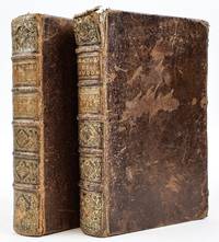
1734rif. 1408797Paris: Guillaume Cavelier 1734. Later Editions. Hardcover. Octavos Four Volumes bound as two. The first book contains a single volume; the second book contains the second volume of Anatomie Chirurgicale in addition to two other standalone works. In Good condition. In full brown leather bindings; spines paneled with elaborate gilt decoration and gilt titling on tan leather labels; gilt roll to board edges. Scuffing and wear to boards with peeling to covers. All edges and fore corners rubbed with sections of exposed board. Chipping to heads and tails of spines; heaviest at head of Vol. I with head band nearly detached. Text block edges show shelf wear and dust soiling. Lightly age toned and very sparsely foxed throughout. Splitting to hinges. Rear pastedown in Vol. I and both pastedowns in Vol. II are separating from the boards. ~2 in. closed tear in Vol. I p. 39. Splitting in gutters of several signatures throughout both volumes due to damage to end bands. Closed tears affect some fold-out plates. Tidemarks affect pages in the rear of both volumes. CONTENTS: Anatomie Chirurgicale Vol. I xxiv 514 2 pages plus plates i-xxx; Anatomie Chirurgicale Vol. II viii 313 336-352 453-465 366-403 pages plus plates xxxi-xlviii bound together with Observations Anatomiques viii 175 pages plus 8 plates and Six Observations 2 36 2 pages. Shelved in Room A. The first work in this set is the second edition of Flemish surgeon Jean Palfin's Jan Palfijn's very famous treatise on anatomy which was first published in 1726 under the title Anatomie du Corps Humain. This work and its earlier edition were very influential in the medical field for their detailed anatomical illustrations and descriptive actionable advice for surgeons becoming a widespread authoritative medical text throughout Europe and other parts of the world. The second work bound together with Vol. II of Anatomie Chirurgicale is a French translation of Frederik Ruysch's Observationum Anatomico-Chirurgicarum Centuria. Ruysch was a renowned anatomist and botanist in his own right most well-known for his anatomical museum - the Cabinet of Curiosities - and his improved embalming technique which preserved bodies for longer periods of time in a more visually life-like state. The final work also bound together with Vol. II of Anatomie Chirurgicale and Observations Anatomiques is a revised edition of Six Observations by Michel Brisseau who is notable for being the first to demonstrate the true nature of cataracts as an increased opacity of the lens of the eye rather than a film that covered the eye which was the prevailing belief at the time. Together these works comprise a collection of highly influential studies treatises and observations on anatomy and surgery by some of the most notable medical practitioners of the late 17th and early 18th centuries. 1408797. Special Collections - Upstairs. Guillaume Cavelier hardcover books
1828rif. 40962London 1828. Parliament. House of Commons. Report from the select committee on anatomy. Folio. 150pp. London: House of Commons 22 July 1828. 331 x 212 mm. 19th century boards rebacked and recornered in calf light edgewear. Very good copy. Old medical library stamp on title and first page. First Edition of this highly interesting and entertaining report on the British body-snatching crisis. Since the mid-eighteenth century obtaining cadavers for teaching purposes had been regulated in Britain by the Murder Act of 1752 which stipulated that only the corpses of executed criminals could be used for dissection. By the beginning of the nineteenth century however improvements in medical science coupled with a substantial drop in the number of executions caused the demand for cadavers to far outstrip the legal supply. This situation was ripe for exploitation by "resurrection men" criminals who robbed the graves of the newly deceased and sold their corpses to teachers of anatomy who of necessity turned a blind eye to the illegality of these transactions. Some grave-robbers even resorted to murder including the infamous William Burke who in December 1828 was arrested in Edinburgh for the murders of over a dozen victims whose corpses he and his partner Hare sold to an anatomical demonstrator connected to Edinburgh University. In the first half of 1828 in response to increasing calls for reform the British Parliament appointed a committee to "enquire into the manner of obtaining subjects for dissection by schools of Anatomy and the State of law affecting persons employed in obtaining and dissecting bodies." During the course of its investigation the committee heard testimony from a wide range of witnesses from eminent medical men to procurers of bodies for medical schools these last identified only by initials. The medical men included Sir Astley Cooper Benjamin Collins Brodie John Abernethy William Lawrence Herbert Mayo Granville Sharp Pattison who himself was indicted for body-snatching at the age of 23 Thomas Southwood Smith Henry Halford John Webster and Benjamin Harrison the treasurer of Guy's Hospital. The witness list can be found on page 13 of the committee's report. The testimony of these men reproduced in full in the report is followed by several appendices including tables of paupers' deaths broken down by parish; the committee was proposing legislation that would allow the state to seize unclaimed corpses from workhouses and sell them to surgical schools. The committee's efforts were successful: In 1832 Parliament passed the Anatomy Act granting licenses to teachers of anatomy and giving physicians surgeons and medical students legal access to corpses unclaimed after death. Wise The Italian Boy: A Tale of Murder and Body Snatching in 1830s London 2004. Garrison-Morton.com 7096. . unknown books
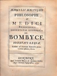
1669rif. 20151London: J Martyn & J Allestry Regie Societatis Typographos 1669. First Edition. Full contemporary calf. Good; some modest overall toning and light spotting to several plates text crisp and clear; binding worn chafed and taped along the backstrip; short clean tears into four plates no loss; very good margins throughout. 4to 10 100 pp 12 engraved folding plates. Contemporary engraved bookplate of the Viscount of Scudmore with a shelf mark in ink. Initial and final blank present. "Malpighi's work on the silkworm represents the first monograph on an invertebrate and records one of the most striking pieces of research work on his part. He dissected the silkworm under the microscope with great skill and observed its intricate structure; before the appearance of this work the silkworm was believed not to have internal organs" G-M #293. "In de Bombyce Malpighi had carefully observed the artisan nature construct each of the three stages - larva chrysalis and moth - through which the silkworm is formed. He further remarked on the specific apparatuses with which the silkworm is provided among them the air ducts tracheae and the blood duct with a number of pulsating centers corcula" DSB IX p.65. Frati #20; Norman #1428; Wing M-349. J Martyn & J Allestry Regie Societatis Typographos unknown books
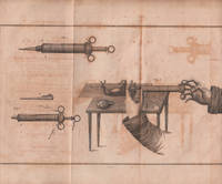
1813rif. 14086Philadelphia: M Thomas 1813. First English Edition. full contemporary calf. Very good; some minor offsetting; joints chafed. 8vo. Folding plate. An elusive and important title. Le Gallois described the action of the vagus nerve on respiration and located the respiratory center in the medulla. The plate accompanying the text is dramatic. See G - M # 928 citing the first French edition; Austin # 1139; Norman # 1324; Clarke & O'Malley Human Brain pp. 294 - 296. M Thomas unknown books
1669rif. 479London: Martyn 1673 & 1669. First editions second issue. Full contemporary calf. Very good; rebacked. 4to. There are 16 engraved plates. The first edition sheets of both works bound up with a new general title. The first work G-M #469 is a milestone in the history of modern embryology and was unsurpassed until Wolf's rare treatise at the end of the 18th century. Malpighi's book on embryology was an original work that presented many new and correct ideas. The second work on the silkworm G-M #293 is the first book dealing with an invertebrate. He developed new dissecting techniques and was responsible for the identification of all the animal's internal structure. In the copy offered here the Epistola to Malpighi from Oldenburg is bound after the title for De Formatione though it belongs with the second treatise. This copy retains the license leaf. Frati p.27; see Adelmann for a lengthy account; G-M # 293 & # 469; Wing M-351; see Norman #1428 & 1429. Martyn unknown books
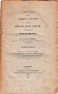
1824rif. 5269Glasgow: Wardlaw & Cunninghame 1824. Second Enlarged Edition UK prtg. Original blue paste paper boards and paper label. Good; spine abraded; covers loose. 8vo 4 adverts dated Feb. 1824 4 xxix 1 - blank 31 - 510 2 - plate list pp. 10 plates. The second enlarged edition edited by Granville Pattison who was professor of anatomy at the Univ. of Maryland in America. Pattison was a student of Burns in Edinburgh. The two men were on friendly terms and after Burns' early death Pattison took over his copyrights business and lectureship at the College Street medical School. This edition is enlarged by appx. 100 pages; probably issued subsequent to the American edition of a year prior when Pattison was still resident in Baltimore. Burns was a noted anatomist. This book is similar to that of Morgagni's great treatise in that it attempts to correlate pathological signs to specific diseases. The illustrations are specific. An important book in cardiology see Bedford's comments in his catalog and most uncommon in the original boards as issued. See G - M # 405 & #3055; Goldschmid p. 113; see the Pattison biography. Wardlaw & Cunninghame hardcover books
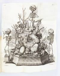
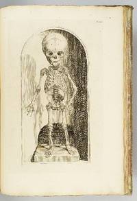
1417rif. ST20185Amsterdam: Joannes Wolters then Jansson-Waesberg 1701-14; 1715-16. FIRST EDITIONS. 224 x 168 mm. 8 3/4 x 6 1/2". 4 p.l. 62 pp. 1 leaf errata; 2 p.l. 98 pp.; 2 p.l. 70 pp.; 1 p.l. 55 pp.; 1 p.l. 54 pp. 2 p.l. 30 pp. 1 leaf 92 pp. 2 leaves 44 pp. 4 leaves 68 pp. 8 leaves 72 pp. 1 leaf errata 12 leaves 78 pp. 10 parts in one volume. Contemporary stiff vellum smooth spine with ink titling yapp edges. WITH 42 ENGRAVED PLATES as called for by Norman six of these folding. Text in Latin and Dutch. Blake 395; Garrison-Morton 389; Heirs of Hippocrates 627; Jeremy Norman's HistoryofMedicine.com 623; Norman 1875. Vellum lightly soiled front pastedown slightly defective but the original unsophisticated binding in perfect order. Final plate with small hole due to adhesion to facing page minor damage done to image a few other trivial imperfections a couple of negligible marginal tears a faint ink stain across the top inch of text a tiny burn hole but AN UNUSUALLY BRIGHT CLEAN COPY INTERNALLY with all the important plates in exceptionally fine condition. Scarce when seen bound from its original 10 parts as here this work describes the memorable "Anatomical Treasure" of Dutch physician and anatomist Frederick Ruysch being illustrated by surrealistic charmingly macabre engravings depicting "fantastic dream-like concoctions constructed of human anatomical parts." History of Medicine Described by Norman as "probably the most original artist in the history of anatomical preparations" Ruysch 1638-1731 "enjoyed making up elaborate three-dimensional emblems of mortality from his specimens" which included fetal skeletons and preserved organs. Heirs to Hippocrates says that "the engraved illustrations deserve special mention for their whimsical almost surrealistic quality: quaintly posed skeletons surrounded by stuffed monsters strange reptiles dried plants and sea creatures." According to Norman "Ruysch's methods allowed him to prepare organs such as the liver and kidneys and keep entire corpses for years. He used a mixture of talc white wax and cinnabar for injecting vessels and an embalming fluid of alcohol made from wine or corn with black pepper added. Using his injection methods Ruysch was the first to demonstrate the occurrence of blood vessels in almost all tissues of the human body thereby destroying the Galenic belief that certain areas of the body had no vascular supply. He was also the first to show that blood vessels display diverse organ-specific patterns." Ruysch's dioramas were displayed in his home museum in Amsterdam which was open to the public; visitors included Tsar Peter the Great of Russia who purchased the collection in 1717 for the foundation of Russia's first public museum the St. Petersburg Kunstkammer. Unfortunately the Russian climate was not kind to the fragile constructions and all deteriorated over time; the dramatic plates here are the best record we have of this unique collection. Joannes Wolters, then Jansson-Waesberg unknown books
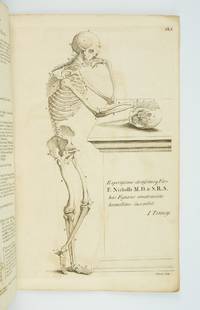
1770rif. ST20219London: Printed for Robert Sayer ca. 1770. 380 x 242 mm. 15 x 9 1/2". 4 leaves of text followed by plates. Original gray paper wrappers. WITH EIGHT FINE ENGRAVED PLATES of the male body in various poses three depicting the skeleton and five the musculature. Russell 816; Wellcome V p. 273; ESTC N51161. See also Russell "John Tinney's Compendium Anatomicum and its publishers." Wrapper a bit soiled and foxed corners torn with two snags two-inch and half-inch to lower cover most of spine chipped away but the stitching holding the book firmly together; mild offsetting from engravings and a few spots of foxing but a really excellent copy internally the leaves fresh and clean and with very good impressions of the engravings. This rare collection of striking anatomical engravings is wonderfully preserved in its original unrestored wrappers. First published in 1743 as "Compendium Anatomicum" the work's plates are adapted from the famous anatomies of Vesalius and Cowper. It was intended as a reference work for artists but as its name suggests it was published with a wider audience in mind. As Tinney humbly tells the reader in the subtitle this is "a work not only very useful but absolutely necessary to painters statuaries and all professors of drawing and design as well as a proper introduction to the study of anatomy for the use of young surgeons" not to mention an "instructive furniture for the studies and libraries of the curious." The work remained in print for a full century though because it was a frequently used book few copies have come down to us in collectible condition. John Tinney ca. 1706-61 was an engraver and print seller who dealt in a wide variety of material being particularly known for maps and satirical prints as well as the present work. Our edition while undated almost certainly dates to the period between 1762-70. The first posthumous edition of "A Compendious Treatise" was released in 1762 by Robert Sayer a map and print seller who was an associate of Tinney's during his lifetime and who likely purchased his stock of plates from his widow following his 1761 death. In 1770 Sayer began a partnership with John Bennett after which the firm was known as "Sayer and Bennett." Since the present edition was published under Sayer's name alone it no doubt comes from the period following the 1762 edition but before the partnership. The present copy has made it through the centuries in remarkably good condition given its flimsy binding and the plates inside with their cheerfully macabre figures remain quite fresh and pleasing. Printed for Robert Sayer unknown books
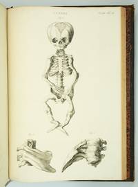
1793rif. ST20586Lugduni Batavorum Leyden: S. & J. Luchtmans 1793 1827 1835. FIRST EDITION A COMPLETE SET. 525 x 350 mm. 20 3/4 x 14". Four volumes. First two volumes in contemporary cat's paw calf volumes III and IV in contemporary flamed calf raised bands spines gilt in compartments with central fleuron one red and one black morocco label. WITH 206 ENGRAVED ANATOMICAL PLATES all with guards. Front pastedowns with bookplate of Harvard Medical School Library; library shelf markings in white at bottom of spines; bookseller's ticket of Schönhof & Mueller Foreign Books Boston. Bibliotheca Walleriana 8549; Choulant pp. 312-13; Larousse XIX vol. 14 p. 171. Lucas L. Boer Peter L. J. Boek Andries J. van Dam and Roelof-Jan Oostra "History and highlights of the teratological collection in the Museum Anatomicum of Leiden University The Netherlands " American Journal of Medical Genetics Part A Volume 176 no. 3 2018 pp. 618-37. Shallow chips across the tail of first two volumes covers variably abraded one noticeably so corners somewhat worn partially with boards showing through vague additional signs of wear but the bindings solid and retaining much of their original appeal; plates in first two volumes with faint overall browning either because of paper stock or because of contact with guards a handful of trivial tears but all in all an extremely pleasing set the plates in volume IV very clean and bright and the plates elsewhere and the wide-margined text clean and fresh throughout. This is an extremely scarce complete set of a very substantially proportioned anatomical work with more than 200 striking giant folio engravings that emphasize birth defects and that range from clinically cheerless to heartbreaking. A physician and professor of anatomy at Leyden Eduard Sandifort 1742-1814 was commissioned by the University and the city council to publish this account of the school's anatomical collections with a focus on pathological anatomy including especially congenital abnormalities. After the publication of the first two volumes in 1793 the third and fourth were issued following Sandifort's death by his son Gerard 1779-1848 who followed in his father's professorial footsteps in Leyden. The first second and fourth volume are illustrated with copperplate engravings which Larousse tell us make this book "one of the most beautiful works of this genre" though "beautiful" needs to be understood here to mean "convincing" or "compelling" rather than aesthetically appealing. The first volume concludes with nine full-size depictions of skulls; the illustrations in the second and fourth are of a more affecting nature consisting of malformed bodies and body parts damaged or destroyed by disease and birth defects. The engravings of the conjoined twins in these volumes are especially powerful--consisting of poignant even haunting images showing undeserving victims of pronounced congenital defects though some are softened with a touch of the fanciful in their presentation. The thoroughness and accuracy of the work is so great that it remains of scholarly value even today. A recent study by Dutch curators and anatomical scholars Lucas L. Boer Peter L. J. Boek Andries J. van Dam and Roelof-Jan Oostra used "Museum Anatomicum" as a guide to identify and re-diagnose the University's historic teratological collections noting that "many historically made diagnoses could not be improved upon after re-diagnosing the specimens with contemporary dysmorphological knowledge actually confirming that these old collectors were perhaps the first dysmorphologists and can be seen as true pioneers in the field of teratology." Although single volumes and incomplete sets of this work come on the market with some regularity we have been able to trace only five complete sets at auction since 1872. And ours especially fresh and wide-margined in pleasing contemporary calf is a particularly attractive specimen. S. & J. Luchtmans unknown books
1739rif. ST12883Leyden: Johann Arnold Langerak 1739. First Edition in Latin. 540 x 380 mm. 21 1/4 x 15". 70 leaves of text.Translated by William Dundass. Original red quarter vellum over marbled boards raised bands UNTRIMMED EDGES. Engraved printer's device on title large decorative initials and tailpieces EXTRA ENGRAVED TITLE AND 114 STRIKING PLATES OF ANATOMICAL FIGURES three folding. Wellcome II 401; Heirs of Hippocrates 468; Choulant-Frank pp. 252-3; Russell 213. ◆Vellum on spine rather worn with three one-and-one-half-inch pieces broken away revealing structure underneath paper boards quite chafed other minor problems externally but an entirely solid unrestored binding. A couple of plates with short closed marginal tears one folding plate with one-inch closed tear into image no loss untrimmed edges a little browned and brittle with isolated small chips occasional minor foxing or insignificant stains three plates lightly browned but still AN UNUSUALLY ATTRACTIVE COPY INTERNALLY with the broadest of margins with especially clean and fresh leaves and with rich impressions of the plates. With plates of notable visual impressiveness this was the finest anatomy book in England during the first half of the 18th century; it also was largely a plagiarism borrowing most of its engraved content from a work by the Dutch physician Govert Bidloo published in 1685. Bidloo's work was the first large-scale anatomical atlas to appear after Vesalius' epoch-making "De Humani Corporis Fabrica" and the plates which are highly praised by Norman and Garrison-Morton are characterized by startlingly detailed life-size depictions of the human body both adult and infant with figures flayed to reveal muscles opened to show organs and unfleshed to exhibit bones. According to Choulant-Frank Bidloo's publishers sold 300 impressions of these plates to Cowper probably to recoup some of their money after disappointing sales. Cowper took Bidloo's original 105 plates added nine of his own and produced an English translation of the original Latin text to accompany them. Discussing the original plates produced by Gerard de Lairesse 1641-1711 Norman says that the figures are displayed "in an emotional almost tender manner contrasting the raw dissected parts with the full soft surfaces of uncut flesh placing flayed bound figures in ordinary nightclothes or bedding setting ordinary household objects such as books jars or cabinets in the same scene as cut-up torsos or limbs. His illustrations brought the qualities of Dutch still-life painting into anatomical illustration and gave a new darker spiritual expression to the significance of the act of dissection." When Cowper's version of the atlas first appeared as "The Anatomy of Humane Bodies" in London in 1698 there was also a 1737 Leyden printing in English before our more scholarly Latin edition Bidloo complained to the Royal Society and accused Cowper of plagiarism and fraud resulting in much acrimony and heated pamphleteering between the two physicians. Notwithstanding this scandal Cowper's achievements and discoveries--including the pair of glands that bear his name--were considerable and his text improved significantly upon the original work. Unfortunately as the DNB notes "the notoriety of this case has served to obscure a true appreciation of Cowper and of his many original contributions to anatomical illustration." The atlases of Bidloo and Cowper appear on the market regularly but at 540 x 380 mm. the present copy is distinguished by its size which is significantly larger than what is typically seen with this edition--we have not been able to trace a copy larger than ours from marketplace or institutional records. Johann Arnold Langerak unknown books
1830rif. D7399Italy c. 1830. Hardcover. Very Good. Modern half morocco and marbled boards gilt-stamped lettering and ornament on spine; 430 x 560 mm; contains 26 anatomical drawings in red and grey chalk most of them full-page but also including double-page spreads tipped onto stubs. Spine tips and corners gently bumped; some faint dampstaining; a few leaves with repaired tears. Images of skeletal and muscular representations of arms legs feet torsos and heads. hardcover books
rif. 15090b13/14. Auflage Urban & Schwarzenberg München 1985. Je 628 Seiten mit zahlreichen Abbildungen Pappbände. - gute Exemplare - unknown
rif. ria9780323756549_inpPaperback / softback. New. New Book; Fast Shipping from UK; Not signed; Not First Edition; N/A paperback
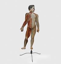
1890rif. 179459Peoria Illinois: American Manikin Company 1890. The bare bones of anatomy An educational model depicting male anatomy. It was positioned as a cheaper and more user-friendly alternative to Louis Auzoux's papier-mâché manikins themselves an accessible innovation from earlier wax models. The American Manikin represents a crucial step in the widening accessibility and popularization of anatomy and is a precursor to the mass-produced teaching models of the 20th century. The model is double-sided one face depicting muscles nerves tendons and the spinal column and the other showing muscles veins and bones. The latter face has alphabetically labelled hooks onto which illustrated steel attachments can be affixed. When all the plates are in place the model can be "dissected" by removing each layer in sequence eventually revealing the skeleton. Several of the attachments have additional diagrams on the reverse; for example the attachment illustrating the nerves of the brain also features a diagram of the inner ear on the other side. First produced in 1888 the manikin was designed by Elias Smith 1828-1913 who also published the Atlas of Colored Plates Illustrating Features of Anatomy 1889. Smith was an enterprising businessman who held patents for bicycle saddles and dental equipment. The accompanying advertisement booklet makes extensive reference to the American Manikin's superiority over Auzoux's French model. The attachment for the model's hand is illustrated with the company's calling card. The manikin was issued with 40 attachments of which 24 are present. An accompanying Teachers' Handbook was also produced not present. Wooden model approximately 1070 x 407 mm chromolithographic illustrations affixed to both faces one face with 27 metal hooks. With 24 steel attachments with similar illustrations metal stand comprising base and three attachable legs. Housed in original wooden case 1020 x 510 mm chromolithographic anatomical diagrams printed on inner panels instructional leaflet affixed to same. With 62-page advertisement booklet. Case with near-contemporary postage label of Lewis Bohnett 1880-1970 antique-collector and proprietor of "Trader Lew's Relic Museum" California; "fragile" label dated 1939 to same. Lightly worn from use lacking single hook cockling and a few chips to chromolithographic overlays colours bright; superficial wear to case: in very good condition standing firm. unknown
rif. ANAIS-1878576488Bryan Edwards Pub Inc. other. Good. 4.8x6.3x6. Buy with confidence. Excellent Customer Service & Return policy. Bryan Edwards Pub Inc unknown
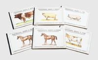
1900rif. 188515London: Vinton & Company Ltd c. 1900. A set of elaborate moveable books depicting the anatomy of farmyard animals. Each includes five layers of folding colour diagrams showing the skeleton arteries and veins muscles and organs accompanied by a key. It was printed in Bavaria a prestigious centre of moveable books and home to skilled artisanal paper engineers such as Ernest Nister and Lothar Meggendorfer. This is the second edition of the set. Six works oblong quarto. Each 8 pp. and with folding chromolithographic illustration comprising two large overslips and multiple lift-the-flap tabs representing internal anatomy. Original printed boards black cloth backstrips wire-stitched as issued. Foxing toning and soiling to boards and contents wear to backstrips wire-stitches rusted but holding firm outer margins of contents with a few chips and damp stains a handful of neat repairs to folding stubs colours bright: in very good condition. hardcover
rif. ria9780443113666_inpPaperback / softback. New. New Book; Fast Shipping from UK; Not signed; Not First Edition; N/A paperback
2009rif. SKU0516066Muze Publishing 2020-09-02. paperback. Good. 8x0x11. Textbook May Have Highlights Notes and/or Underlining BOOK ONLY-NO ACCESS CODE NO CD Ships with Tracking Muze Publishing paperback
2009rif. SKU0608653Muze Publishing 2020-09-02. paperback. New. 8x0x11. New Textbook Ships with Tracking Muze Publishing paperback
rif. ria9781455776849_inpPaperback / softback. New. New Book; Fast Shipping from UK; Not signed; Not First Edition; N/A paperback
rif. ria9780702079269_inpPaperback / softback. New. New Book; Fast Shipping from UK; Not signed; Not First Edition; N/A paperback
rif. ria9781455776832_inpCards. New. New Book; Fast Shipping from UK; Not signed; Not First Edition; N/A unknown
rif. A9781345346527Hardback. New. hardcover
rif. A9781346173856Hardback. New. hardcover
2020rif. x-1914207238Muze Publishing 2020. Paperback. New. 132 pages. German language. 11.00x8.50x0.30 inches. Muze Publishing paperback