2 498 résultats
1879réf. 5450Paris, Librairie Germer Baillière et Cie, 1879 ; in-8, broché ; 112 (numérotées 329 à 448), 8 pp. de catalogue.
réf. 14259o.J. Pathologe. 1900 Prof. in Straßburg, 1906 Prof. u. Direktor am Pathologischen Institut der Medizinischen Klinik in Düsseldorf, 1907 in Zürich, 1911 in Marburg, von 1913-35 Prof. in Würzburg. 1863 Leipzig - 1949 Mittenwald. Brustbild mit Brille. Photogravure um 1905. 300 x 200 mm.
1949réf. 218330Basel, Karger, 1949-61. M. Abb. Bibl.-Hlwd. Teils St. u. Sign. a. Tit.
1936In 8? (cm 27), Leg. tutta tela, pagg.862 con numerose ill. b.n. n.t., segni del tempo alla cop., buon es. Prima ed unica edizione. Supplemento al vol. VI dell'"Archivio Italiano di anatomia e istologia patologica". Alberto Pepere (Napoli, 1873 - Genova, 1940) fu illustre anatomista e istologo-patologo
1961réf. 704764Stuttgart, Thieme, 1961. 4°. M. 818 Abb. X, 808 S. Hlwd. Sign. a. Tit.
réf. 44809ABJena Verl. v. G. Fischer. Achte, umgearb. und vermeh. Aufl. Erster Band: Infektionskrankheiten 8° 784 S.: mit 278 Abb im Text und 14 farb. Taf. OLwd. Vord. Deck. gebrochen. ansonst gut. Zust. 8
1963réf. 704770Stuttgart, Thieme, 1963. M. mehr. Tab. u. 128 (teils farb.) Abb. XI, 390 S. OLwd.
1870réf. 628016Wien, Braumüller, 1870. M. 2 lith. Taf. u. 4 Textholzschn. OU. angestaubt.
1897réf. 188865London, 1860-1897. M. einigen (teils farb.) Taf. u. Abb. OLwd. M. Goldpräg. M. kl. Rsign. Einbde. etwas berieben. St. a. Tit. Bei 11 zusätzl. Einbd. etwas fleckig u. Vors.teils angeschmutzt.
réf. 22909o.J. Arzt, Gerichtsmediziner, Pharmakologe. 1680 Garnisonsarzt der Festung Philippsburg, 1687 Prof. in Gießen, 1728 kaiserl. Leibarzt. Mitglied der Leopoldina. 1657 Gießen - 1729 ebd.. Fast Halbfigur im Schriftoval, unten Sockel mit latein. Vers. Kupferstich von E. Nunzer. 171 x 134 mm.
réf. 29358o.J. Mediziner, Pathologe. 1762 Prof. der allgemeinen Pathologie und pathologischen Anatomie in Leipzig. Übernahm 1877 den Lehrstuhl für spezielle Pathologie u. Therapie in Leipzig, Direktor der Medizinischen Klinik. Entdeckte die Fettembolie in der Lunge. 1829 Dehlitz /Saale - 1888 Leipzig. Brustbild nach halbrechts. Stahlstich von Weger. 215 x 155 mm.
réf. 13335o.J. österr. Pathologe. 1875 Prosektor am Garnisonsspital Nr. 1, 1882 an den Krankenanstalten Rudolfstiftung, 1885 Prof. der pathologischen Histologie u. Bakteriologie an der Universität Wien, leistete Pionierarbeit auf dem Gebiet der Lungenpathologie, wies als erster im Leichenblut Tuberkelbazillen nach, entdeckte den Erreger der Genickstarre, Gründer der ersten Lungenheilstätte Alland in Österreich. 1845 Schiltern /NÖ - 1920 Wien. Brustbild nach halblinks. Photogravure bei Eckstein um 1910. 305 x 200 mm.
1921réf. 243580Berlin, Hirschwald, 1905-1921. M. 354 Taf. (teils lith. u. farb.), 661 Curven u. 471 Textabb. Hldr. m. Rsign. St. a. Vorsatz, Tit. u. mehr. S. Ecken teilw. bestoßen. Einige Rücken eingerissen u. teilw. lose. Einige S. braunfl. u. teilw. beschmutzt.
réf. 22845o.J. Mediziner, Kliniker. 1863 Prof. d. Pathologie u. Therapie sowie Direktor der Medizinischen Klinik in Erlangen, 1874 Direktor des Allgemeinen Krankenhauses in München, errichtete 1877 das erste deutsche klinische Institut. 1829 Greifswald - 1902 München. Brustbild im Profil nach rechts. Photogravure um 1905. 305 x 205 mm.
1990Verona, Cartiere del Garda, 1990, 4to brossura, pp. 40 con ill. a colori n.t.
1906In 8, pp. XI + 922 + (2b). Br. ed. Ponderosa pubblicazione contenente notizie relative a tutte le zone e i comuni dichiarati malarici presentati in ordine alfabetico. Sono presenti, fra le altre, le province di Alessandria, Aquila, Ascoli Piceno, Avellino, Bari, Belluno, Benevento, Bergamo, Bologna, Brescia, Cagliari, Caltanissetta, Campobasso, caserta, Catania, Catanzaro, Chieti, Como, Cosenza, Cremona, Ferrara, Foggia,... Napoli, Novara, Padova, Palermo..., Siena, Torino, ecc...
1895réf. 142509Wien, Alfred Hölder k. u. k. Hof- und Universitäts-Buchhändler 1895, bzw. 1904, 1895, 1906, 1897 1895. 431 S., Abb. 25*17 cm. OHalblederband mit Farbschnitt.
réf. 9954de 6 numéros du Progrès Médical, consacrés à Xavier Bichat (1771-1802), célébrissime médecin anatomiste et physiologiste, né à Thoirette alors en Bresse (aujourd'hui dans le Jura), élevé à Poncin dans le Bugey puis à Nantua et Lyon.
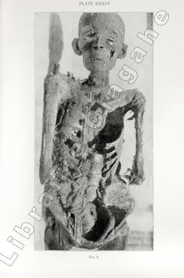
réf. 18329Edited by Roy L. Moodie. Chicago, Illinois, University of Chicago Press, 1921 ; in-8, cartonnage d’éditeur de percaline vert foncé, titre doré au dos, XX, 372 pp., photographie assis en frontispice et 71 planches hors-texte dont 8 en couleurs.
2014réf. 2090502113709472Not Available 2014. Soft Cover. Fine. The book is in fine condition. Not Available paperback
réf. 51731P., Jouve, 1917, in 8° broché, 251 pages, non coupé.
1691réf. 24166Lyon Thomas AMAULRY 1691 -in-8 plein-veau 1 fort volume, reliure plein veau brun in-octavo (binding full calfskin in-octavo), RELIURE DE L'EPOQUE, dos à nerfs (spine with raised bands), titre frappé "or", pièce de titre sur fond bordeaux avec un filet "or" en encadrement, tomaisons frappées "or" avec une roulette "or" de part et d'autre dans un encadrement d'un filet "or", filet à froid de part et d'autre des nerfs, entre-nerfs a fleuron "or" en losange dans un encadrement d'un filet "or" avec rinceaux "or" aux angles, roulette large "or" en pied, trés léger manque de cuir à la coiffe de tête et petit manque à la coiffe de pied, roulette "or" sur les coupes avec manques de dorure, toutes tranches lisses jaspées rouges, orné d'une vignette en bas du titre gravée sur bois en noir + Une planche hors-texte de description de Microbes (p.480) (MANQUANTE), [(10) + 740 + 15 p. de Tables] Pages, 1691 Lyon Thomas AMAULRY Editeur,
1699réf. 24165Lyon Thomas AMAULRY 1699 -in-8 plein-veau 2 forts volumes, reliure plein veau brun in-octavo (binding full calfskin in-octavo), RELIURE DE L'EPOQUE, dos à nerfs (spine with raised bands), titre frappé "or", pièce de titre sur fond bordeaux avec un filet "or" en encadrement, tomaisons frapées "or" avec une roulette "or" de part et d'autre dans un encadrement d'un filet "or", filet à froid de part et d'autre des nerfs, entre-nerfs a fleuron "or" en losange dans un encadrement d'un filet "or" avec rinceaux "or" aux angles, roulette large "or" en pied, manque de cuir à la coiffe de tête et petit manque à la coiffe de pied du Tome II, tache brune sur les plats du Tome I, coins écornés, roulette "or" sur les coupes avec manques de dorure, toutes tranches lisses jaspées rouges, sans illustrations (no illustration) excepté la marque en bas du titre de l'Editeur Thomas AMAULRY gravée sur bois en noir, [(6) + 722 + 62 p. de Tables] + [(6) + 718 + 66 p. de Tables] Pages, 1699 Lyon Thomas AMAULRY Editeur,
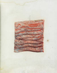
1789réf. 120a new era for Anatomical Pathology: The raise of coloured illustrations First edition of Bleuland's histological investigation of duodenal tissue Bleuland Jan.Icon tunicæ villosæ intestini duodeni juxta felicem vasculorum impletionem. Ipsis coloribus qui in præparato conspiciuntur edita. Trajecti ad Rhenum Utrecht: apud B. Wild et J. Altheer 1789. 4to 21 x 19 cm later paper wrapper pp. 10 2-last leaf blank missing signature Aâ´ B² B2 blank missing 1 coloured plate representing a sample of duodenal tissue Jan Bleuland 1756-1838 was famous physician professor of Anatomy Physiology Obstetrics and rector of Utrecht University. He became famous perfecting a technique of injecting fixatives and dyes into human body soft tissues structures to reveal previously invisible anatomical details. The printed illustrations of the results of his technique are unique in their precision in depicting microscopic information. Among Bleuland's greatest achievements was his ability to visualize vascular systems making even the smallest blood vessels perfectly visible preserving in the same time the true colours and texture of the tissues. The possibility to obtain detailed and realistic tissue images developed and improved by Bleuland togheter with the work of his colleague the Scottish pathologist Matthew Baillie 1761 – 1823 gave a significant boost to the innovation of anatomical pathology. During his brilliant career he carried out more than two thousand preparations and his immense collection of anatomical specimens is now preserved at the Utrecht Medical University. "Bleuland focused on the soft parts of human body which involved a much wider spectrum of organs and diseases and posed a different set of problems to technicians in the preparation of the specimens" Meli p.211 op. cit. "During his career Bleuland published a number of illustrated anatomical works printed in color showing a special interest in the fine structure of healthy and diseased states of the organs and tunics of the digestive tract. Bleuland made a collection of over two and a half thousand anatomical preparations with the assistance of Petrus Koning whom he had trained as his attendant and as a surgeon from 1800" Meli op.cit. Conditions: small restoration at lower margins in general a good copy. Provenance: Ownership signature at title page. References: Domenico Bertoloni Meli The Rise of Pathological Illustrations: Baillie Bleuland and Their Collections Bulletin of the History of Medicine Volume 89 Number 2 Summer 2015 pp. 209-242 Article; Florian Rodari Anatomie de la couleur 124; James G. Mundies's Cabinet of Curiosite's February 2008; Blake p. 50; not in Waller or Wellcome. B. Wild et J. Altheer
First edition, small 8vo (180 x 120 mm), 64pp., light soiling and spotting, new endpapers, recent calf-backed marbled boards, red morocco spine label. First published in 1772 with Perry using the pseudonym William Adams for the first four editions.