5.838 resultados
1739ref. ST12883Leyden: Johann Arnold Langerak 1739. First Edition in Latin. 540 x 380 mm. 21 1/4 x 15". 70 leaves of text.Translated by William Dundass. Original red quarter vellum over marbled boards raised bands UNTRIMMED EDGES. Engraved printer's device on title large decorative initials and tailpieces EXTRA ENGRAVED TITLE AND 114 STRIKING PLATES OF ANATOMICAL FIGURES three folding. Wellcome II 401; Heirs of Hippocrates 468; Choulant-Frank pp. 252-3; Russell 213. ◆Vellum on spine rather worn with three one-and-one-half-inch pieces broken away revealing structure underneath paper boards quite chafed other minor problems externally but an entirely solid unrestored binding. A couple of plates with short closed marginal tears one folding plate with one-inch closed tear into image no loss untrimmed edges a little browned and brittle with isolated small chips occasional minor foxing or insignificant stains three plates lightly browned but still AN UNUSUALLY ATTRACTIVE COPY INTERNALLY with the broadest of margins with especially clean and fresh leaves and with rich impressions of the plates. With plates of notable visual impressiveness this was the finest anatomy book in England during the first half of the 18th century; it also was largely a plagiarism borrowing most of its engraved content from a work by the Dutch physician Govert Bidloo published in 1685. Bidloo's work was the first large-scale anatomical atlas to appear after Vesalius' epoch-making "De Humani Corporis Fabrica" and the plates which are highly praised by Norman and Garrison-Morton are characterized by startlingly detailed life-size depictions of the human body both adult and infant with figures flayed to reveal muscles opened to show organs and unfleshed to exhibit bones. According to Choulant-Frank Bidloo's publishers sold 300 impressions of these plates to Cowper probably to recoup some of their money after disappointing sales. Cowper took Bidloo's original 105 plates added nine of his own and produced an English translation of the original Latin text to accompany them. Discussing the original plates produced by Gerard de Lairesse 1641-1711 Norman says that the figures are displayed "in an emotional almost tender manner contrasting the raw dissected parts with the full soft surfaces of uncut flesh placing flayed bound figures in ordinary nightclothes or bedding setting ordinary household objects such as books jars or cabinets in the same scene as cut-up torsos or limbs. His illustrations brought the qualities of Dutch still-life painting into anatomical illustration and gave a new darker spiritual expression to the significance of the act of dissection." When Cowper's version of the atlas first appeared as "The Anatomy of Humane Bodies" in London in 1698 there was also a 1737 Leyden printing in English before our more scholarly Latin edition Bidloo complained to the Royal Society and accused Cowper of plagiarism and fraud resulting in much acrimony and heated pamphleteering between the two physicians. Notwithstanding this scandal Cowper's achievements and discoveries--including the pair of glands that bear his name--were considerable and his text improved significantly upon the original work. Unfortunately as the DNB notes "the notoriety of this case has served to obscure a true appreciation of Cowper and of his many original contributions to anatomical illustration." The atlases of Bidloo and Cowper appear on the market regularly but at 540 x 380 mm. the present copy is distinguished by its size which is significantly larger than what is typically seen with this edition--we have not been able to trace a copy larger than ours from marketplace or institutional records. Johann Arnold Langerak unknown books
1823Questa straordinaria opera di anatomia è composta da: - un vol. di testo in folio (mm. 510x330), mz. pelle mod. con ang., dorso a cordoni con tit. oro: “Explication des planches anatomiques du corps humain.. par le Docteur F. Antonmarchi”, pp. (6),VII, V-VIII,288. - una raccolta di tavole in folio max. (mm. 635x410) contenuta in un solido astuccio (spacchi ai bordi) in mz. pelle mod. con legacci, dorso a cordoni con tit. oro. Come da dettagliata descriz. nel vol. di testo, le tavole - tutte a doppia pag. - sono così ripartite: 48 sono in chiaroscuro e 35 sono ripetute al tratto, ciasc. con una o più figure, disegnate da Pedretti e pregevolmente litografate; l’importante collezione è introdotta da un frontespizio inquadrato in cornice floreale (sempre a doppia pag.). "Edizione originale", molto rara trovarla completa. “Antonmarchi served as personal physician to Napoleon at St. Helena and achieved a certain editorial notoriety as the editor of Paolo Mascagni’s posthumous "Prodromo della grande anatomia". Upon termination of his obligations with Mascagni’s heirs, he seems to have considered the rights his own and republished the plates in the present edition, with some modifications, under his own name - without denying their provenance. He nevertheless failed to cite Mascagni on the title page and this was poorly received by the medical community. The work was conceived so that the entire human body might be represented (in life size) by joining three of the plates together. This may be achieved a number of times to depict the complete skeleton, the blood vessels, the muscles (front and back) and the viscera. Further detailed plates are dedicated to the brain, the heart, the lymphatic system, etc.”. Cfr. Choulant “History and bibliography of anatomic illustration”, pp. 319-320 che cita erroneam. 45 + 45 tavv. - Hirsch,I, p. 154-55 con 83 tavv. come il ns. esemplare. Il vol. di testo: occhietto restaur. per strappi, alone al marg. super., fiorit. o arross. interc. nel t. - Le tavv.: prime 12 con alone al marg. inf., altre 20 ca. più o meno fiorite, ma certam. un buon esemplare.
1725ref. 5551Leiden, J. du Vivie et J. & H. Verbeek, 1725. 2 Bde. 21 Bll., 572 S.; 4 Bll.; S. 577 - 684, 1 Bl., S. 685 - 1156, 26 Bll. mit 2 wdh. Kupfer-Titelvignetten und 38 Textkupfern. 1 Kupfertitel und 82 (12 gefalt.) Kupfertafeln (es fehlt das Kupfer-Porträt). Gr.-Fol. Mod. HLdr. auf 5 Bünden mit Rückentitel und modernen Buntpapierbezügen ((etw. berieben). [6 Warenabbildungen]
1600in-folio (mm.300x200, ff.12 nn., pp.442, ff.14 nn., titolo inciso con figure allegoriche e ritratto dell'autore, al recto del f.di dedica ritratto di Enrico IV con le figure della Giustizia e della Prudenza; 26 stupende figure anatomiche, assai dettagliate, inc . a piena pagina, gran numero di capilettera istoriati in silografia su otto linee d'altezza. Testo in elegante carattere tondo e corsivo. Legatura coeva in pieno cuoio bruno, dorso a nervi con titolo e ricchi fregi oro, in perfetto stato. Seconda edizione (prima Paris 1599). E' uno dei più importanti trattati anatomici e medici del tardo cinquecento e le belle figure si ispirano ai rami di Vesalio, Valverde, Coiter ed altri. Il Du Laurens (1558-1609) fu medico di corte di Enrico IV e Maria de Medici e successivamente capo della prestigiosa facoltà di medicina a Montpellier. Per primo, tra l'altro, descrisse con precisione l'arteria polmonare e la circolazione del sangue in generale. Esemplare assai bello, puro e fresco.. Choulant p.222. Cushing n.117. Durling 1312. Wellcome 1917. Bird 774..
ref. 4394P., BAILLIERE, 1837-1841, 3 VOLUMES in 8 de TEXTE et 1 ATLAS GRAND IN FOLIO D'ATLAS, reliés en demi-basane rouge dos ornés de fers de filets dorés,T.1 (texte): 16pp. (compris faux-titre+titre), 638pp., 6 PLANCHES hors texte, T.2 (texte) : (2-faux titre + titre), 620pp., T.3 (texte) : (2-faux-titre + titre) 810pp. - ATLAS : 3 feuillets non chiffrés (faux titre+titre+dédicace), 4pp. (table), 43 feuillets non chiffrés d'explication des planches (soit 86pp. au total), 60 MAGNIFIQUES PLANCHES EN COULEURS
1793ref. ST20586Lugduni Batavorum Leyden: S. & J. Luchtmans 1793 1827 1835. FIRST EDITION A COMPLETE SET. 525 x 350 mm. 20 3/4 x 14". Four volumes. First two volumes in contemporary cat's paw calf volumes III and IV in contemporary flamed calf raised bands spines gilt in compartments with central fleuron one red and one black morocco label. WITH 206 ENGRAVED ANATOMICAL PLATES all with guards. Front pastedowns with bookplate of Harvard Medical School Library; library shelf markings in white at bottom of spines; bookseller's ticket of Schönhof & Mueller Foreign Books Boston. Bibliotheca Walleriana 8549; Choulant pp. 312-13; Larousse XIX vol. 14 p. 171. Lucas L. Boer Peter L. J. Boek Andries J. van Dam and Roelof-Jan Oostra "History and highlights of the teratological collection in the Museum Anatomicum of Leiden University The Netherlands " American Journal of Medical Genetics Part A Volume 176 no. 3 2018 pp. 618-37. Shallow chips across the tail of first two volumes covers variably abraded one noticeably so corners somewhat worn partially with boards showing through vague additional signs of wear but the bindings solid and retaining much of their original appeal; plates in first two volumes with faint overall browning either because of paper stock or because of contact with guards a handful of trivial tears but all in all an extremely pleasing set the plates in volume IV very clean and bright and the plates elsewhere and the wide-margined text clean and fresh throughout. This is an extremely scarce complete set of a very substantially proportioned anatomical work with more than 200 striking giant folio engravings that emphasize birth defects and that range from clinically cheerless to heartbreaking. A physician and professor of anatomy at Leyden Eduard Sandifort 1742-1814 was commissioned by the University and the city council to publish this account of the school's anatomical collections with a focus on pathological anatomy including especially congenital abnormalities. After the publication of the first two volumes in 1793 the third and fourth were issued following Sandifort's death by his son Gerard 1779-1848 who followed in his father's professorial footsteps in Leyden. The first second and fourth volume are illustrated with copperplate engravings which Larousse tell us make this book "one of the most beautiful works of this genre" though "beautiful" needs to be understood here to mean "convincing" or "compelling" rather than aesthetically appealing. The first volume concludes with nine full-size depictions of skulls; the illustrations in the second and fourth are of a more affecting nature consisting of malformed bodies and body parts damaged or destroyed by disease and birth defects. The engravings of the conjoined twins in these volumes are especially powerful--consisting of poignant even haunting images showing undeserving victims of pronounced congenital defects though some are softened with a touch of the fanciful in their presentation. The thoroughness and accuracy of the work is so great that it remains of scholarly value even today. A recent study by Dutch curators and anatomical scholars Lucas L. Boer Peter L. J. Boek Andries J. van Dam and Roelof-Jan Oostra used "Museum Anatomicum" as a guide to identify and re-diagnose the University's historic teratological collections noting that "many historically made diagnoses could not be improved upon after re-diagnosing the specimens with contemporary dysmorphological knowledge actually confirming that these old collectors were perhaps the first dysmorphologists and can be seen as true pioneers in the field of teratology." Although single volumes and incomplete sets of this work come on the market with some regularity we have been able to trace only five complete sets at auction since 1872. And ours especially fresh and wide-margined in pleasing contemporary calf is a particularly attractive specimen. S. & J. Luchtmans unknown
1793ref. ST20586Lugduni Batavorum Leyden: S. & J. Luchtmans 1793 1827 1835. FIRST EDITION A COMPLETE SET. 525 x 350 mm. 20 3/4 x 14". Four volumes. First two volumes in contemporary cat's paw calf volumes III and IV in contemporary flamed calf raised bands spines gilt in compartments with central fleuron one red and one black morocco label. WITH 206 ENGRAVED ANATOMICAL PLATES all with guards. Front pastedowns with bookplate of Harvard Medical School Library; library shelf markings in white at bottom of spines; bookseller's ticket of Schönhof & Mueller Foreign Books Boston. Bibliotheca Walleriana 8549; Choulant pp. 312-13; Larousse XIX vol. 14 p. 171. Lucas L. Boer Peter L. J. Boek Andries J. van Dam and Roelof-Jan Oostra "History and highlights of the teratological collection in the Museum Anatomicum of Leiden University The Netherlands " American Journal of Medical Genetics Part A Volume 176 no. 3 2018 pp. 618-37. Shallow chips across the tail of first two volumes covers variably abraded one noticeably so corners somewhat worn partially with boards showing through vague additional signs of wear but the bindings solid and retaining much of their original appeal; plates in first two volumes with faint overall browning either because of paper stock or because of contact with guards a handful of trivial tears but all in all an extremely pleasing set the plates in volume IV very clean and bright and the plates elsewhere and the wide-margined text clean and fresh throughout. This is an extremely scarce complete set of a very substantially proportioned anatomical work with more than 200 striking giant folio engravings that emphasize birth defects and that range from clinically cheerless to heartbreaking. A physician and professor of anatomy at Leyden Eduard Sandifort 1742-1814 was commissioned by the University and the city council to publish this account of the school's anatomical collections with a focus on pathological anatomy including especially congenital abnormalities. After the publication of the first two volumes in 1793 the third and fourth were issued following Sandifort's death by his son Gerard 1779-1848 who followed in his father's professorial footsteps in Leyden. The first second and fourth volume are illustrated with copperplate engravings which Larousse tell us make this book "one of the most beautiful works of this genre" though "beautiful" needs to be understood here to mean "convincing" or "compelling" rather than aesthetically appealing. The first volume concludes with nine full-size depictions of skulls; the illustrations in the second and fourth are of a more affecting nature consisting of malformed bodies and body parts damaged or destroyed by disease and birth defects. The engravings of the conjoined twins in these volumes are especially powerful--consisting of poignant even haunting images showing undeserving victims of pronounced congenital defects though some are softened with a touch of the fanciful in their presentation. The thoroughness and accuracy of the work is so great that it remains of scholarly value even today. A recent study by Dutch curators and anatomical scholars Lucas L. Boer Peter L. J. Boek Andries J. van Dam and Roelof-Jan Oostra used "Museum Anatomicum" as a guide to identify and re-diagnose the University's historic teratological collections noting that "many historically made diagnoses could not be improved upon after re-diagnosing the specimens with contemporary dysmorphological knowledge actually confirming that these old collectors were perhaps the first dysmorphologists and can be seen as true pioneers in the field of teratology." Although single volumes and incomplete sets of this work come on the market with some regularity we have been able to trace only five complete sets at auction since 1872. And ours especially fresh and wide-margined in pleasing contemporary calf is a particularly attractive specimen. S. & J. Luchtmans unknown books
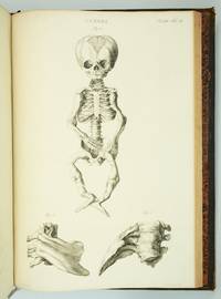
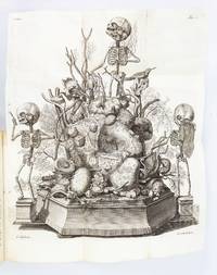
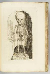
1417ref. ST20185Amsterdam: Joannes Wolters then Jansson-Waesberg 1701-14; 1715-16. FIRST EDITIONS. 224 x 168 mm. 8 3/4 x 6 1/2". 4 p.l. 62 pp. 1 leaf errata; 2 p.l. 98 pp.; 2 p.l. 70 pp.; 1 p.l. 55 pp.; 1 p.l. 54 pp. 2 p.l. 30 pp. 1 leaf 92 pp. 2 leaves 44 pp. 4 leaves 68 pp. 8 leaves 72 pp. 1 leaf errata 12 leaves 78 pp. 10 parts in one volume. Contemporary stiff vellum smooth spine with ink titling yapp edges. WITH 42 ENGRAVED PLATES as called for by Norman six of these folding. Text in Latin and Dutch. Blake 395; Garrison-Morton 389; Heirs of Hippocrates 627; Jeremy Norman's HistoryofMedicine.com 623; Norman 1875. Vellum lightly soiled front pastedown slightly defective but the original unsophisticated binding in perfect order. Final plate with small hole due to adhesion to facing page minor damage done to image a few other trivial imperfections a couple of negligible marginal tears a faint ink stain across the top inch of text a tiny burn hole but AN UNUSUALLY BRIGHT CLEAN COPY INTERNALLY with all the important plates in exceptionally fine condition. Scarce when seen bound from its original 10 parts as here this work describes the memorable "Anatomical Treasure" of Dutch physician and anatomist Frederick Ruysch being illustrated by surrealistic charmingly macabre engravings depicting "fantastic dream-like concoctions constructed of human anatomical parts." History of Medicine Described by Norman as "probably the most original artist in the history of anatomical preparations" Ruysch 1638-1731 "enjoyed making up elaborate three-dimensional emblems of mortality from his specimens" which included fetal skeletons and preserved organs. Heirs to Hippocrates says that "the engraved illustrations deserve special mention for their whimsical almost surrealistic quality: quaintly posed skeletons surrounded by stuffed monsters strange reptiles dried plants and sea creatures." According to Norman "Ruysch's methods allowed him to prepare organs such as the liver and kidneys and keep entire corpses for years. He used a mixture of talc white wax and cinnabar for injecting vessels and an embalming fluid of alcohol made from wine or corn with black pepper added. Using his injection methods Ruysch was the first to demonstrate the occurrence of blood vessels in almost all tissues of the human body thereby destroying the Galenic belief that certain areas of the body had no vascular supply. He was also the first to show that blood vessels display diverse organ-specific patterns." Ruysch's dioramas were displayed in his home museum in Amsterdam which was open to the public; visitors included Tsar Peter the Great of Russia who purchased the collection in 1717 for the foundation of Russia's first public museum the St. Petersburg Kunstkammer. Unfortunately the Russian climate was not kind to the fragile constructions and all deteriorated over time; the dramatic plates here are the best record we have of this unique collection. Joannes Wolters, then Jansson-Waesberg unknown
1417ref. ST20185Amsterdam: Joannes Wolters then Jansson-Waesberg 1701-14; 1715-16. FIRST EDITIONS. 224 x 168 mm. 8 3/4 x 6 1/2". 4 p.l. 62 pp. 1 leaf errata; 2 p.l. 98 pp.; 2 p.l. 70 pp.; 1 p.l. 55 pp.; 1 p.l. 54 pp. 2 p.l. 30 pp. 1 leaf 92 pp. 2 leaves 44 pp. 4 leaves 68 pp. 8 leaves 72 pp. 1 leaf errata 12 leaves 78 pp. 10 parts in one volume. Contemporary stiff vellum smooth spine with ink titling yapp edges. WITH 42 ENGRAVED PLATES as called for by Norman six of these folding. Text in Latin and Dutch. Blake 395; Garrison-Morton 389; Heirs of Hippocrates 627; Jeremy Norman's HistoryofMedicine.com 623; Norman 1875. Vellum lightly soiled front pastedown slightly defective but the original unsophisticated binding in perfect order. Final plate with small hole due to adhesion to facing page minor damage done to image a few other trivial imperfections a couple of negligible marginal tears a faint ink stain across the top inch of text a tiny burn hole but AN UNUSUALLY BRIGHT CLEAN COPY INTERNALLY with all the important plates in exceptionally fine condition. Scarce when seen bound from its original 10 parts as here this work describes the memorable "Anatomical Treasure" of Dutch physician and anatomist Frederick Ruysch being illustrated by surrealistic charmingly macabre engravings depicting "fantastic dream-like concoctions constructed of human anatomical parts." History of Medicine Described by Norman as "probably the most original artist in the history of anatomical preparations" Ruysch 1638-1731 "enjoyed making up elaborate three-dimensional emblems of mortality from his specimens" which included fetal skeletons and preserved organs. Heirs to Hippocrates says that "the engraved illustrations deserve special mention for their whimsical almost surrealistic quality: quaintly posed skeletons surrounded by stuffed monsters strange reptiles dried plants and sea creatures." According to Norman "Ruysch's methods allowed him to prepare organs such as the liver and kidneys and keep entire corpses for years. He used a mixture of talc white wax and cinnabar for injecting vessels and an embalming fluid of alcohol made from wine or corn with black pepper added. Using his injection methods Ruysch was the first to demonstrate the occurrence of blood vessels in almost all tissues of the human body thereby destroying the Galenic belief that certain areas of the body had no vascular supply. He was also the first to show that blood vessels display diverse organ-specific patterns." Ruysch's dioramas were displayed in his home museum in Amsterdam which was open to the public; visitors included Tsar Peter the Great of Russia who purchased the collection in 1717 for the foundation of Russia's first public museum the St. Petersburg Kunstkammer. Unfortunately the Russian climate was not kind to the fragile constructions and all deteriorated over time; the dramatic plates here are the best record we have of this unique collection. Joannes Wolters, then Jansson-Waesberg unknown books
ref. 1403Lugduni batavorum, Joannem Arnoldum Langerak, 1739, un fort volume grand in folio (535x365mm,leaf 525x355mm) relié en PLEIN MAROQUIN ROUGE, (reliure de l'époque), dos orné de fers dorés, toutes tranches dorées, filets dorés sur les plats, 9 ff. (titre gravé, titre imprimé en noir en en rouge, dedicace...), 59 ff. (explication des planches), 2 ff (index), 114 PLANCHES (105 pl. numérotées 1 à 105 + 9 planches numérotées 1 à 9
ref. 482Rome, Antonii de Rubeis, 1741, un volume in folio, (42,20 cm x 30 cm) relié en plein parchemin, dos muet (habiles restaurations sur les plats, quelques traces de poussière dans les marges de la dernière page de texte), 2 feuillets non chiffrés (titre+avis au lecteur), 84pp., 27 SUPERBES PLANCHES
ref. 13967Traité pratique du dessin et de la peinture Tome V Ecole française. In 12 broché, couverture illustrée, 120x190mm, faux –titre, titre, VII, 460 pages, 204 illustrations dans le texte, Paris Librairie Garnier frères, sans date. Couverture doublée, dos renforcé. Intérieur, très bon état
1990ref. ART5932MBroché, 343 pages, paru le 1er décembre 1990 chez RMN, très bon état général.